
Kidney Cross Section Anatomy
|
|---|
- Left suprarenal vein
- Left gonadal vein
- Portion of the left ascending lumbar vein
- The segmentation of the renal arteries allows for independent blood flow to segments of the kidney, which is why surgeons can do a resection without compromising the function of the entire kidney.
- Junction of the renal pelvis and ureter
- Pelvic brim
- Passage through the wall of the bladder

The kidneys lie in the retroperitoneal space on the posterior abdominal wall on either side of the T12-L3 vertebrae. The right kidney usually lies more inferior than the left kidney because of the presence of the liver on the right side. The right kidney is separated from the liver to create the hepatorenal recess (aka. Morison's pouch).
The renal veins form from the confluence of the interlobar veins, and empty into the inferior vena cava (IVC), which empties back into the right side of the heart. The left renal vein is longer than the right (due to the position of the IVC on the right side of the body) and also drains venous blood from the:
The renal arteries arise from the abdominal aorta at the level of L1/L2 and lie anterior to the renal veins. The right renal artery is longer than the left renal artery (due to the position of the aorta on the left side of the body). They divide into the interlobar arteries, and then further segment into cortical arteries, eventually becoming afferent arterioles. Afferent arterioles flow into the glomeruli where filtration occurs.
This is a flat, funnel shaped extension of the superior aspect of the ureter and the apex of the renal pelvis is continuous with the ureter. It goes on to divide into 2-3 major calices and then further into minor calices.
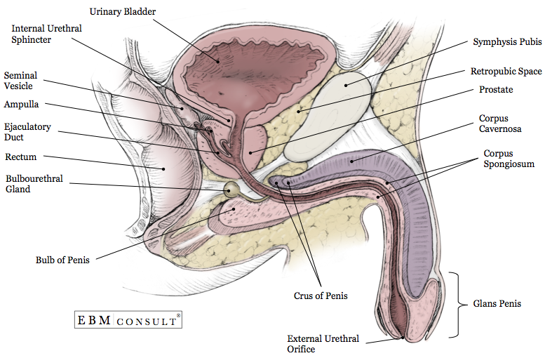
The ureter is about 25-30 cm long and at its superior end collects the final urine filtrate from the minor and major calyces. The ureter carries the urine filtrate inferiorly, passes over the pelvic brim (location where the common iliac arteries bifurcate) and then drains into the bladder. There are 3 places where the ureter is more constricted, which serve as common spots for renal stones to obstruct:
Keywords
|
|---|
|