
Dens (Odontoid) Fractures - General Review
Summary:
-
A
fracture of the odontoid bone (also called the dens), is an upward extension of
C2 cervical vertebrae (i.e., axis) up into the C1 cervical vertebrae (i.e., atlas) and is held in place partially by the alar, apical and transverse
ligaments.
- There are three types of dens fractures with type II being the most common. Types II & III are commonly treated with a halo vest or surgical fixation.
- If starting out with basic radiographs, you would assess by doing an open mouth or odontoid view. If an abnormality is present, a CT or MRI of the head and c-spine can more definitively determine the type of dens fracture.
Dens (Odontoid) Fractures
|
|---|
- Type I = An oblique avulsion of the tip of the dens (treated with cervical collar)
- Type II = Fracture at the base of the dens in a transverse plane (most common type; a non-union would be treated with a halo vest or surgical fixation).
- Type III = Fracture extends down into the body of C2 and is considered unstable (treated with halo or anterior screw fixation)
- Age
- Stability of the patient
- Mechanism of injury
- Presence of other traumatic injuries or neurologic deficits.
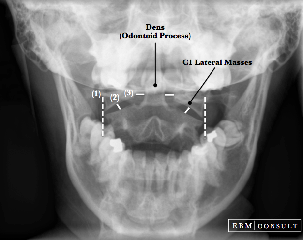
- Open Mouth (Odontoid view) Radiograph: Reveals asymmetry of the spaces between the dens (located on C2 and projecting up) and the lateral masses of C1 (atlas). The lateral masses usually line up with respect to the margins of C2 (axis).
- CT & MRI Scans: If there are neurologic deficits present or the mechanism of injury is concerning, then a CT scan of the head and neck along with a CT angiogram of the neck to evaluate for vertebral artery injuries or spasm. If there are contraindications to a CT scan or IV contrast, then an MRI can be done to provide more definitive interpretation of where the fracture line occurs in the dens.
 Fracture Image")
There are 3 Types of Dens Fractures:
When assessing for the presence of a dens fracture, the choice of initial imaging is influenced by the:
In general radiographs (such as the odontoid view) would be reserved for younger patients, who are not unstable, do not need more advanced imaging (such as a CT or MRI) in the same anatomical areas for evaluation of other injuries.
Imaging Modalities:
To learn more about how to read cervical spine radiographs using various radiographic lines and processes for interpretation select an item below. Many of the same radiographic methods of interpretation for assessing the presence of a cervical spine injury can be utilized across basic radiographs, CT scans or MRIs.
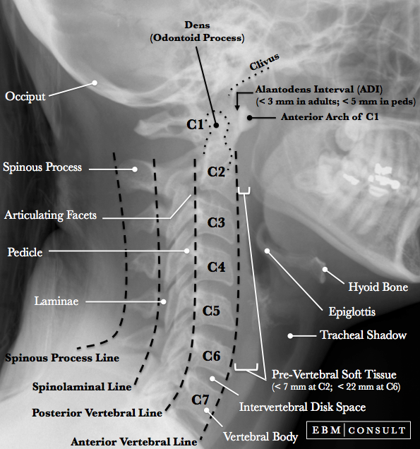
Lateral C-Spine Odontoid View SAC & Torg Ratio