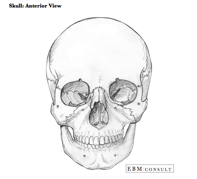
Anatomy: Skull Anterior View
Summary:
- The skull (cranium) is divided into 2 main functional parts: neurocranium and viscerocranium.
- The major anterior skull bones include: Frontal bone, Mandible, Maxilla, Nasal bone, Parietal bone, Temporal bone, and Zygomatic bone.
- The foramen of the anterior skull include (top to bottom): Supraorbital foramen, Zygomatic foramen, Infraorbital foramen, and Mental foramen. Nerves exit out of the foramen.
Note About Images: Scroll over the image to see the labels.
Anatomy: Skull Anterior View
|
|---|
- Neurocranium:
- Bony part that covers the brain and its membranous coverings, the cranial meninges, proximal portions of the cranial nerves, and vasculature of the brain.
- Made up by 8 bones (ethmoidal, frontal, occipital, sphenoidal & the 2 sets of parietal and temporal bones)
- Viscerocranium:
- Represents the bones of the anterior part of the skull Made up by 15 bones (ethmoid, mandible, & vomer along with 2 sets of maxilla, nasal, inferior nasal conchae, lacrimal, palatine and zygomatic bones
- Calvaria:
- Formed by the flat bones (frontal, temporal and parietal and are united by a fibrous interlocking sutures). Note: During early childhood the sphenoid and occipital bones are united by hyaline cartilage known as synchondroses.
- Pneumatized Bones:
- These are bones with air cells or sinus which are thought to reduce to the weight of the head and include the ethmoid, frontal, temporal, and sphenoid. The ethmoid and sphenoid are known to contain the sinuses.
- Supraorbital foramen
- Zygomatic foramen
- Infraorbital foramen
- Mental foramen
- Malar Rash = The is the rash seen on the skin of the face that is distributed in the area overlying the zygomatic bones in patients with SLE (systemic lupus erythematosus). It gets this name because the zygomatic bones were once called the malar bones.
- Mental Foramen = Extraction of the teeth can result in the bone to resorb in that area. If all of the teeth are extracted in the area of mental foramen it can sometimes disappear and cause damage to the nerve exiting the foramen. If patients have dentures on this can put additional pressure on the nerve and thus cause pain with chewing. In addition, if all of the teeth are extracted in the mandible this can decrease the vertical dimensions of the face causing an overclosure effect.
- Le Fort Fractures = This is a fracture involving the maxilla and is broken down into 3 different variations.
- Supraorbital Margin & Supracillary Arches = The supracilliary arches make up the bone rim just above the supraorbital margin by forming a sharper rim. When this area receives a direct injury the skin will commonly lacerate.
Skull Anterior View Image
Note: Scroll over or tap on the image to see the labels.

Note: Scroll over or tap on the image to see the labels.
Bones of the Anterior Skull
The skull (cranium) is divided into 2 main sections:
Other important aspects of the skull include:
Foramen of the Skull
The following foramen are seen on the anterior skull view:
Nerves exit out of the foramen.
Translation to Clinical Practice
There are a few clinical applications related to cranial (or skull) anatomy that are important to consider:

