Open Mouth Odontoid Radiograph
Summary:
The open mouth odontoid radiograph (x-ray) is used to assess for the presence of an upper cervical spine injury. Common injuries to the upper cervical spine include:
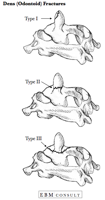
- Dens Fracture (i.e., C2 Odontoid Fracture)
- Jefferson's Fracture (i.e., C1 Burst Fracture)
- Transverse Ligament Injury
- Basilar Invagination
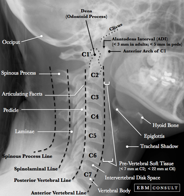
Being able to read and recognize common landmarks when interpreting an open mouth odontoid view is important in determining who has an unstable condition that may warrant more aggressive intervention. Below are some common things to consider when reading these radiographic images.
Note About Images: Scroll or place cursor over the images below to see the landmarks and labels you need to be able to identify.
Editors:
- Anthony J. Busti, MD, PharmD, FNLA, FAHA
- Dylan Kellogg, MD
Last Updated: August 2015
Open Mouth Odontoid Xray
|
|---|
- Make sure the lateral masses of C1 (atlas) do not hang over the lateral masses of C2 (axis).
- The rule of Spence would suggest that if there is more than a combined (total of both sides) overhang of 6.9 mm or more of the lateral masses of C1 in relation to the C2 lateral masses then there is concern for an injury to the transverse ligament and an MRI should be done. While this radiographic rule can be used, it is important to recognize that it may not always correlate well and management decisions should not be made without first obtaining an MRI. Furthermore, an MRI is preferred over a CT scan, since the CT scan may not be able to show the maximal positions of displacement in the fractures.
- Make sure there is no asymmetry of the articular spaces between the lateral masses of C1 and the body of C2 (axis).
- Make sure there is no asymmetry of the articular spaces between the dens and the lateral masses of C1
-
If the lateral masses of C1 extend out beyond the C2 lateral masses this would be most concerning for a Jefferson (or Burst) fracture of C1.
-
If there is any asymmetry between the articular spaces (mainly lines 3) this would be most concerning for either a Jefferson (or Burst) fracture of C1 or a dens fracture (or Odontoid fracture) of C2 or possible transverse ligament injury.
- This is a craniocervical junction abnormality where the tip of the dens project up into the foramen magnum.
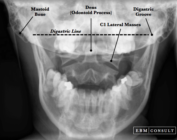
- This is a line connecting the right and left
digastric grooves on a coronal cut of a CT scan or AP skull radiograph.
- It is used to measure the distance from the
tip of the dens (odontoid process) to help evaluate the presence of a basilar
invagination.
- The tip of the dens should be around 11-12 mm below this line.
-
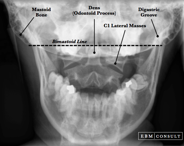
This is a line connecting the tip of the right and
left mastoid bones.
- The distance below
this line to the tip of the dens is used to assess for the presence of basilar
invagination.
- The tip of the dens should be less than 10 mm above this line.
- Make sure there is no overlap of the dens (odontoid process) by the teeth
- Make sure the lateral masses
of C1 (atlas) are symmetrically aligned with lateral masses of C2
- Make sure there is no asymmetry of the articular spaces between the dens and the lateral masses of C1
- Make sure there is no asymmetry of the articular spaces between the lateral masses of C1 and the body of C2 (axis)
Note: Scroll over or tap over image to see lines & labels.

Note: Scroll over or tap over image to see lines & labels.
Line 1
Line 2
Line 3
Clinical Relevance
Note: Scroll over or tap on the image to see the lines & labels

Scroll over or tap on the image to see the lines & labels
Basilar Invagination
Diagastric Line
Bimastoid Line
When reading any radiograph the clinician should establish a process or order they follow each time. While in no particular order, consider evaluating the following:
Note: The presence of a dens fracture, Jefferson fracture, or transverse ligament injury should be give considerable consideration if there is any asymmetry of the articular spaces between the dens and lateral masses or lateral masses and the body of C2.