Digastric Line on Radiographic Image
Summary:
-
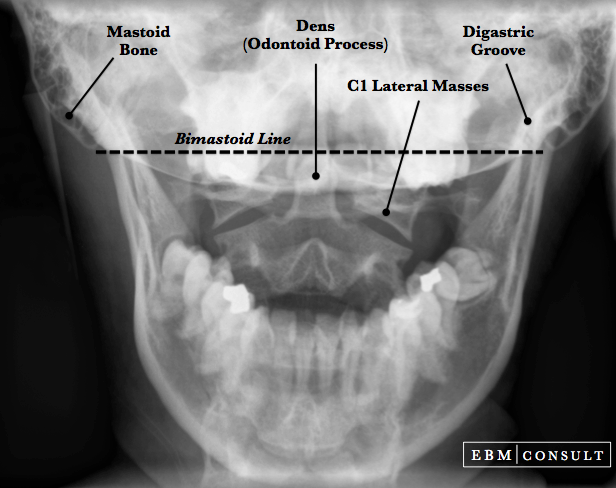
The digastric line is drawn by connecting a line from the right and left
digastric grooves on a coronal cut of a CT scan or AP skull radiograph.
- It is used to measure the distance from the
tip of the dens (odontoid process) to help evaluate the presence of a basilar
invagination (a craniocervical junction abnormality where the tip of the dens
project up into the foramen magnum).
- The tip of the dens should be around 11-12 mm below this line.
Digastric Line
|
|---|