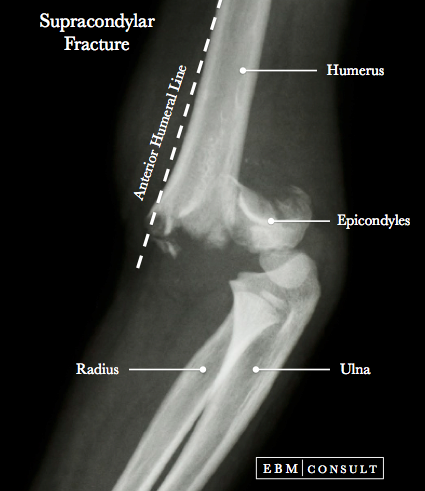
Supracondylar Fracture
Summary:
- A fracture involving the distal aspect of the humerus and just above the epicondyles.
- There types of fractures based on Gartland classification that are influenced by the degree of displacement and degree of bone cortex still intact with type I being managed most commonly with cast immbolization whereas type II & III requiring surgical correction.
- Complications most commonly include nerve palsies (anterior interosseous nerve neurapraxia (most common) and then median nerve, radial nerv, & ulnar nerve palsy).
Supracondylar Fracture - General Review
|
|---|
- Occurs most commonly in pediatric patients
- Injury is most commonly from a fall on outstretched hand (FOOH)
- Type I = Fracture is not displaced
or minimally displaced.
- Treatment: Cast immobilization (if an extension type of
fracture then consider initially splinting into a 20 degree elbow flexion
- Type II = Fracture is
displaced but the posterior cortex is still intact (thus making it a
"greenstick" fracture)
- Treatment: Surgical reduction and correction of fracture
(open reduction, especially vascular compromise is of concern) or percutaneous
pin fixation (mainly 2 lateral pins to avoid injury to ulnar nerve)
- Type III = Fracture is completely
displaced with no bone cortex intact. The distal fragment of the humerus is
displaced posteriorly which can penetrate into the brachialis muscle
- Treatment = Surgical reduction and corrected of fracture (open reduction, especially vascular compromise is of concern or a difficult closed reduction) or percutaneous pin fixation
- Anterior interosseous nerve
neurapraxia (a branch of the median nerve):
- Most common nerve injury from
a supracondylar fracture, especially if there is posterolateral displacement of
the fracture
- Exam Finding = Unable to flex the
interphalangeal joint of the thumb and distal interphalangeal joint of the
index finger, thereby unable to make the "OK" sign.
- Median Nerve Palsy:
- Consider presence of a
compartment syndrome as a median nerve palsy can mask it
- Radial Nerve Palsy:
- Second most common injury,
especially with posteromedial displacement of fracture
- Exam Finding = Unable to extend
the wrist or fingers
- Ulnar Nerve Palsy:
- Seen with flexion type
fractures
- Can result in paralysis or
motor weakness of the muscles of the hand (hypothenar muscles, interosseous
muscles, the deep half of the flexor pollicis brevis, palmaris brevis, and
adductor pollicis) which can cause a "claw-like" deformity (also called the
Duchenne sign)
- Other manifestations include: decreased pinch and grip strength, lateral movement of the fingers and integration of the PIP and MIP joints for flexion. Can also see abduction of the little finger (Wartenberg's sign)
- Lateral views: Displacement of the anterior humeral line, humerotrochlear line,
- AP view of the elbow: Change in Baumann Angle, Medial Epicondylar Epiphyseal (MEE) Angle
- Radiographic Lines of the Elbow: Anterior Humeral Line, Humerotrochlear Angle, Radiocapitellar Line, Baumann's Angle, Medial Epicondylar Epiphyseal Angle
- Injuries of the Elbow: Monteggia Fracture
- Gartland JJ. Management of supracondylar fractures of the
humerus in children. Surg Gynecol Obstet 1959;109:145-154.
- Culp RW et al. Neural injuries
associated with supracondylar fractures of the humerus in children. J Bone
Joint Surg 1990;72A:1211-1215.
- Mallo G et al. Use of the Gartland classification system for treatment of pediatric supracondylar humerus fractures. Orthopedics 2010;33(1):19.
X-Ray Image of Supracondylar Fracture

Note: The above plain radiographic image demonstrates a type III supracondylar fracture since there is complete displacement and no intact bone cortex.
Mechanism of Injury
Classification & Treatment
Supracondylar fractures are classified into 3 main types based on the Gartland system (can be either extension or flexion type):
Complications
Imaging Considerations
Radiograph of the both injured and contralateral elbow (note: the contralateral may be helpful in determining the normal anatomy in some cases). Consider application of the following radiographic lines for evaluation:
Related Content
To see other related content please select from an item below:
References

