McRae Line on Radiographic Image
Summary:
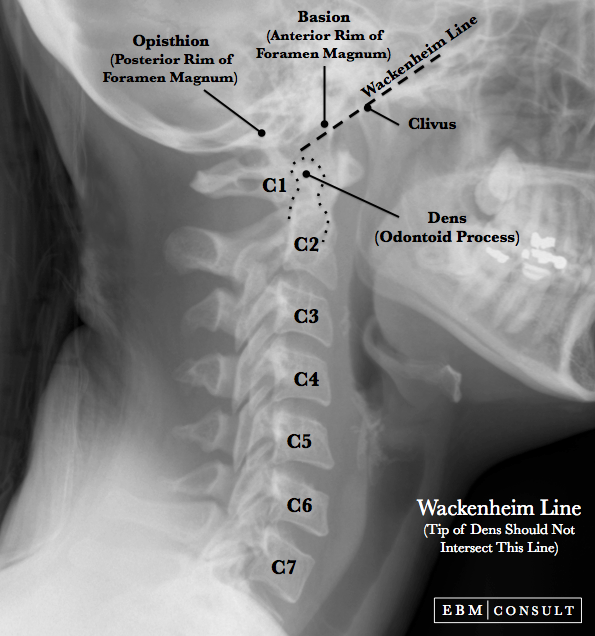
- The McRae line is a line drawn on a lateral radiograph of the skull or on a sagittal cut from a CT or MRI scan that connects the posterior (opisthion) and anterior (basion) aspects of the foramen magnum
- The tip of the dens (or odontoid process) should be ~5 mm below this line. If it is above this line it is concerning for a possible basilar invagination.
McRae Line
|
|---|
Radiographic Image

Note: If the tip of the dens is eroded then the
Redlund-Johnell and modified Ranawat methods (normal CT values for men is >
23.7 mm and for women is > 24.2 mm)
Related Content
To see other related content please select an item below:
Chamberlain Line Wackenheim Line Lateral X-Ray Odontoid View
