AP Neck Radiograph (X-Ray)
Summary:
-
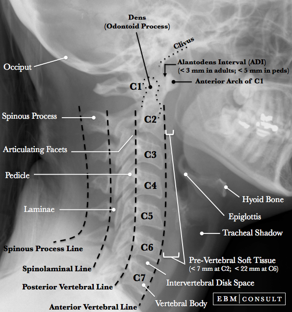
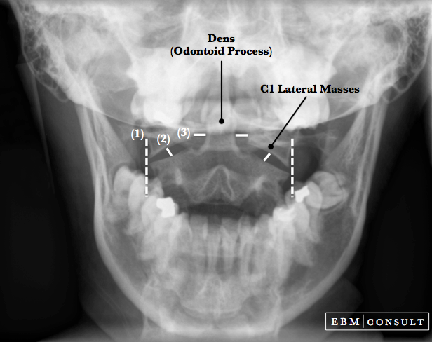
The AP view of the cervical spine (i.e., "c-spine") is one of 3 primary views used to evaluate for the presence of a cervical fracture or subluxation after a traumatic injury involving the head and/or neck.
- This page is dedicated to help you to learn the common anatomical landmarks and a process for how to read an AP neck x-ray.
Note About Images: Scroll over or tap on the image to see the labels and lines.
AP Neck Radiograph
|
|---|
- Do the spinous process appear midline and evenly spaced?
- Do the heights of the vertebral bodies appear to be the same?
- Are the disk spaces uniform in height?
- Are all of the ucinate processes in proper locations and apparent symmetry?
Radiographic AP Neck Image
Note: Scroll over or tap on the image to see the labels & lines

Note: Scroll over or tap on the image to see the labels & lines
How to Read
When reading any radiograph the clinician should establish a process or order they follow each time. While in no particular order, consider evaluating the following: